

CLICK THE LINK
Auto-Refraction
Auto-refraction is an advanced technique that uses the latest technology to evaluate a patient’s corrective prescription. The patient’s face is placed in front of the auto-refractor and a beam of light is painlessly passed over the eyes. This computerized instrument can then quickly and accurately determine the prescription needed for corrective lenses.
Auto-refraction is especially helpful to obtain precise results in certain populations of patients traditionally harder to measure due to an inability to provide responses indicating when their vision appears most clear. These may include young children and patients of any age with:
- Macular degeneration
- Amblyopia or strabismus
- Astigmatism
- Extreme nearsightedness or farsightedness
- Hearing impairment
- Language difficulties
Corneal Topography
The cornea is the clear covering of the front of the eye that bends, or refracts, light rays as they enter the eye. For clear vision to occur, the cornea must have the correct shape and clarity to focus incoming light rays precisely on the retina at the back of the eye.
A computerized test called corneal topography can map out the surface of the cornea, alerting your doctor to the presence of inflammation, scarring or astigmatism. It provides information about the surface power, thickness and shape of the cornea, allowing certain corneal diseases or abnormalities to be diagnosed at a much earlier stage than usual. Corneal topography is a valuable tool prior to a contact lens fitting, vision correction procedure or corneal transplant.
Endothelial Cell Count
Endothelial cell count is a measurement of the number of endothelial cells, which line the inner surface of the cornea. It can also be used to evaluate the size and shape of the endothelial cells. This is important information because the endothelial layer helps maintain the thinness and clarity of the cornea. Once cells are lost, they do not grow back.
Testing of the endothelial cell count is often performed prior to a corneal or cataract surgery. It can help to ensure that the endothelium is healthy enough to for a procedure to be effective. In addition, a low endothelial cell count can indicate the presence of certain diseases, such as Fuchs’ dystrophy.
Eye Ultrasound: A-Scans & B-Scans
Ocular ultrasound is a form of testing employing high-frequency sound waves to create an image of some portion of the eye. It is performed in the ophthalmologist’s office. After anesthetic drops are applied to the eye, the doctor will position the ultrasound wand so it is touching the eye. The ultrasound will generally be completed in 15 minutes and patients do not experience any pain.
A-Scan
A-scan is an ocular ultrasound that provides information on the length of an eye, which is an important factor in determining the selection of an intraocular lens after cataract removal. An A-scan may also be used to diagnose a variety of common eye conditions as well as analyze the shape and size of a mass within the eye.
B-Scan
B-scan is a high-frequency ocular ultrasound that produces a cross-sectional image of the eye and the space behind the eye. The B-scan enables eye care professionals to accurately diagnose and evaluate a wide range of eye conditions including:
- Cataracts
- Retinal detachment
- Tissue damage
- Inflammation
- Vitreous hemorrhage
- Tumor
- Suspected foreign body
- Eye trauma
- Eye pain
- Lens dislocation
Fluorescein Angiography
Fluorescein angiography is a diagnostic procedure used to evaluate the blood vessels in the following parts of the eye:
- Retina
- Choroid
- Optic disc
- Iris
The fluorescein angiography provides doctors with information about the retina. It can also be used to provide information as to the status of current treatments. A fluorescein angiography helps to diagnose and track problems such as:
- Diabetic retinopathy
- Macular degeneration
- Abnormal vessel growth
- Swelling
- Leaking
- Retinal detachment
- Cancer
- Tumors
- Retinitis pigmentosa
Fundus Photography
A fundus photograph is a specialized form of medical imaging. Using a customized camera with high-powered lenses that are mounted to a microscope, photographs are taken of the back of the eye by focusing light through the cornea, pupil and lens. Fundus photographs are used to identify or monitor a wide variety of ophthalmic conditions.
To begin the process, the pupil is dilated with eye drops. The patient will be asked to stare at a fixed device, keeping the eyes focused and still. There will be a series of flashes of light. The process usually takes no more than 10 minutes.
Some of the ophthalmic conditions fundus photography is used for include:
- Glaucoma
- Diabetic retinopathy
- Macular edema
- Microaneurysm
- Optic nerve
Fundus photography has also been used to interpret the results of a fluorescein angiogram.
The Heidelberg Retinal Tomograph (HRT)
The high eye pressure associated with glaucoma can damage your optic nerve before you begin to experience any vision loss. The Heidelberg Retinal Tomograph (HRT) can digitally perceive damage that may indicate the onset of glaucoma, allowing treatment to begin before vision is lost. Similar to MRI (Magnetic Resonance Imaging) and CT (Computed Tomography), HRT is a non-invasive procedure that scans the eye – all you see is a series of flashing red lights.
The Humphrey Visual Field (HVF)
The Humphrey Visual Field is a special automated procedure used to perform perimetry, a test that measures the entire area of peripheral vision that can be seen while the eye is focused on a central point. Patients with glaucoma will often undergo this test on a regular basis in order to determine how quickly the disease is progressing. The Humphrey Visual Field test can also be used to detect conditions within the optic nerve of the eye, and certain neurological conditions as well.
Optical Coherence Tomography (OCT)
Optical coherence tomography (OCT) is an advanced technology used to produce cross-sectional images of the retina, the light-sensitive lining on the back of the eye where light rays focus to produce vision. These images can help with the detection and treatment of serious eye condition such as macular holes, macular swelling, and optic nerve damage.
Pachymetry
Pachymetry is a diagnostic test that is performed to help determine whether a patient has glaucoma. The pachymeter device is used to assess the thickness of the cornea. After anesthetic eye drops are applied, the doctor will place the tip of the pachymeter on the front of the eye.
If the cornea is particularly thin, an intraocular pressure reading may be inaccurately low, and if the cornea is particularly thick, it may result in a reading that is inaccurately high. This knowledge allows the doctor to make adjustments to the pressure reading. A finding of a thin cornea may also indicate an increased risk for the development of glaucoma.
TearLab’s™ Osmolarity Testing
![]() TearLab’s osmolarity testing is an advanced method of diagnosing and treating dry eye conditions. Dry eye is a common disorder that occurs when the eyes are insufficiently moisturized, causing stinging, sensitivity to light, blurred vision and eye fatigue. The eyes may become dry and irritated because the tear ducts don’t produce enough tears, or because of a chemical imbalance in the tears. If left untreated, dry eye can lead to complications including ulcers or scars on the cornea and loss of vision.
TearLab’s osmolarity testing is an advanced method of diagnosing and treating dry eye conditions. Dry eye is a common disorder that occurs when the eyes are insufficiently moisturized, causing stinging, sensitivity to light, blurred vision and eye fatigue. The eyes may become dry and irritated because the tear ducts don’t produce enough tears, or because of a chemical imbalance in the tears. If left untreated, dry eye can lead to complications including ulcers or scars on the cornea and loss of vision.
If dry eye is suspected, the doctor will ask questions about the symptoms and initiate testing. TearLab’s osmolarity testing involves collecting a small sample of the tear fluid from each of the patient’s eyes. The sample is used to measure the salt content, or osmolarity, of the tears. The collection process causes no discomfort and is complete in less than one minute. The evaluation is immediate, providing the doctor with a number that can indicate the presence of dry eye. This measurement helps determine the severity of the condition, allowing the doctor to plan appropriate treatment.
Visual Evoked Potential
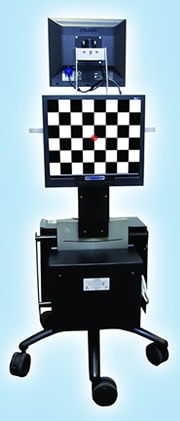 Visual Evoked Potential, commonly referred to as VEP, is a test performed to diagnose a number of conditions, including glaucoma, amblyopia, stroke, brain injury, and multiple sclerosis. The test consists of a blinking checkerboard pattern on a television screen, done to stimulate the visual pathways of the brain. Electrodes placed on the scalp are used to record the patient’s electrical responses, helping to diagnose problems within the optic nerve.
The Diopsys NOVA® system is an advanced VEP testing device that enables the doctor to assess the entire visual pathway from the eye’s lens all the way to the region of the brain responsible for visual processing. This provides a much more complete analysis than standard vision tests, which cannot provide information beyond what can be observed within the eye.
Patients feel no discomfort during the testing and the eyes do not need to be dilated. This testing is safe and can be performed on patients six months old or older to identify a neuro-visual disorder or evaluate the effectiveness of a treatment method. It is particularly valuable for very young children and those with verbal difficulties as no communication is required during Diopsys NOVA testing.
The eyes may be tested together or a patch may be used to test each eye separately. The length of time to complete testing will vary depending upon the amount of information that needs to be compiled.